 |
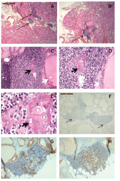
| Figure 1: The thymic tissue was situated at contact to the thyroid (A,B: arrows, images from multistep sections). Thyroid vesicles were at close proximity to thymic lymphoid and Hassall’s corpuscles (without fibrous tissue interposition) (C,D,E: black arrow/Hassall’s corpuscle; white arrow/thyroid follicles). p63 was positive in dispersed intrathymic cells (F, arrows). Cytokeratin AE1/AE3 was expressed by sparse cells in a reticular or small-foci pattern, while CD5 in several lymphocytes. Hematoxilin and eosin stain (A-E), p63 (F), cytokeratin CKAE1/AE3 (G) and CD5 (H) immunohistochemistries; original magnification X2.5(A,B), X5(F), X10(C ), X20(G,H), X40(D), X100(E). |