|
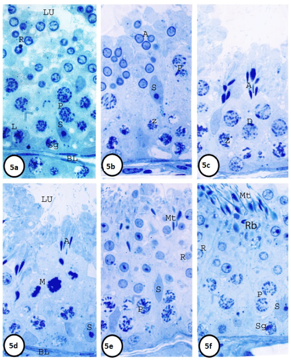
| Figure 5: 5a: Stage I: is characterized by two generations of primary spermatocytes (leptotene and pachytene) and one generation of cap-phase round spermatids 5b: Stage II: is characterized by the leptotene primary spermatocytes were changing to zygotene, and one generation of acrosomal –phase round spermatids. 9c: Stage III: Pachytenes entered the diplotene stage that had larger nuclei (Figure 5c). Spermatids nuclei became more elongated and condensed (acrosomal phase) and arranged in bundles. . In the basal compartment, zygotene spc are additionally seen. 9d: In Stage IV: The most prominent feature of this stage was presence of second meiotic division (M) and secondary spermatocytes (SpII). Zygotene spc (Z) are also identified. 5e: In Stage V-VII, the seminiferous epithelium was composing of two generations of spermatids are observed; elongated and round While only one generation of spc (pachytene, p) is basally located. 5f: In Stage VIII, the maturation phase spermatids on the free border of the seminiferous epithelium .Residual bodies were also demonstrating in this stage and pachytene is basally located. 5a-5f: Semithin sections in the testis of mature donkey (2 years old) showing six cellular associations representing stages I, II, III, IV, V-VII and VIII of the semniferous epithelial cycle, respectively. (Toluidine blue, 1000X). |