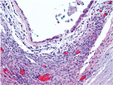
Figure 1:
Mucinous cystadenoma showing a cuboidal epithelium lining and underlying stroma of elongated cells. (Hematoxylin and eosin; 100X). Spindle stromal cells between arrows.