 |
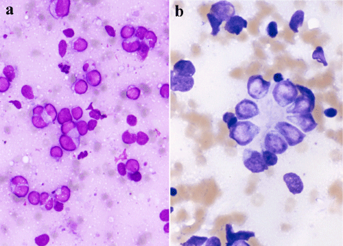
| Figure 6: a) Poorly differentiated neuroblastoma, Giemsa, 40X. Tumor cells possessed bare nuclei with round to oval shape. These nuclei show anisokaryosis and irregular nuclear membrane. b) Poorly differentiated neuroblastoma, Giemsa, 100X. Nuclear arrangements and shapes are clearly observed in Giemsa stain. |