 |
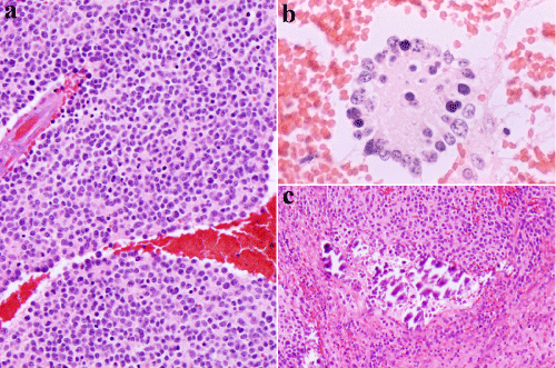
| Figure 2: a) Poorly differentiated neuroblastoma, HE, 40X. Neuroblastic cells have round to oval nuclei in shape. The neuropils are barely seen.b) Poorly differentiated neuroblastoma, HE, 40X. Tumor cells are possessed bare nuclei with round and oval in shape. Homer-Wright rosette arrangements are formed by tumor cells that are radially arranged in circle. The neuropil are stained pink with eosin dye. c) Poorly differentiated neuroblastoma, HE, 40X. Neuroblastic cells are seen in the background. Psammoma bodies are seen occasionally. |