|
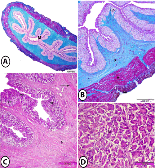
| Figure 2: Histological structure of the fundic region. 2A: the mucosa (M) was thrown into many longitudinal folds (Crossmon's trichrome). 2B: the fundic region showing lamina propria (Lp), submucosa (S) and serosa (se) stained green, while, tunica muscularis (M) stained red. (Crossmon's trichrome). 2C: the folded mucosa (m) of fundic region includes epithelium (ep) and lamina propria (Lp) that contains fundic glands (fg), surrounded with submucosa (S) that contains diffuse smooth muscle bundles (arrow). (Haematoxylin and Eosin). 2D: fundic glands (fg) resemble the pancreatic acini. No parietal or chief cells. (Haematoxylin and Eosin). |