 |
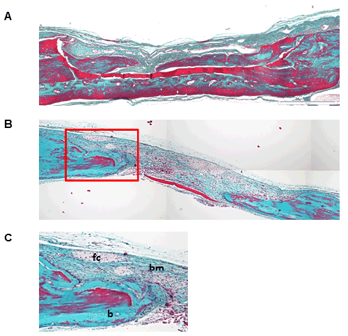
| Figure 2: Masson Goldner trichrome staining of a calvarial crosssection embedded in paraffin after 4 weeks of healing. (A) untreated control lesion, (B) oxycellulose treated bone lesion, magnification x20; (C) higher magnification of the highlighted area (red frame), magnification x100. b=bone; bm=bone marrow / connective tissue; fc=foam cells. |