|
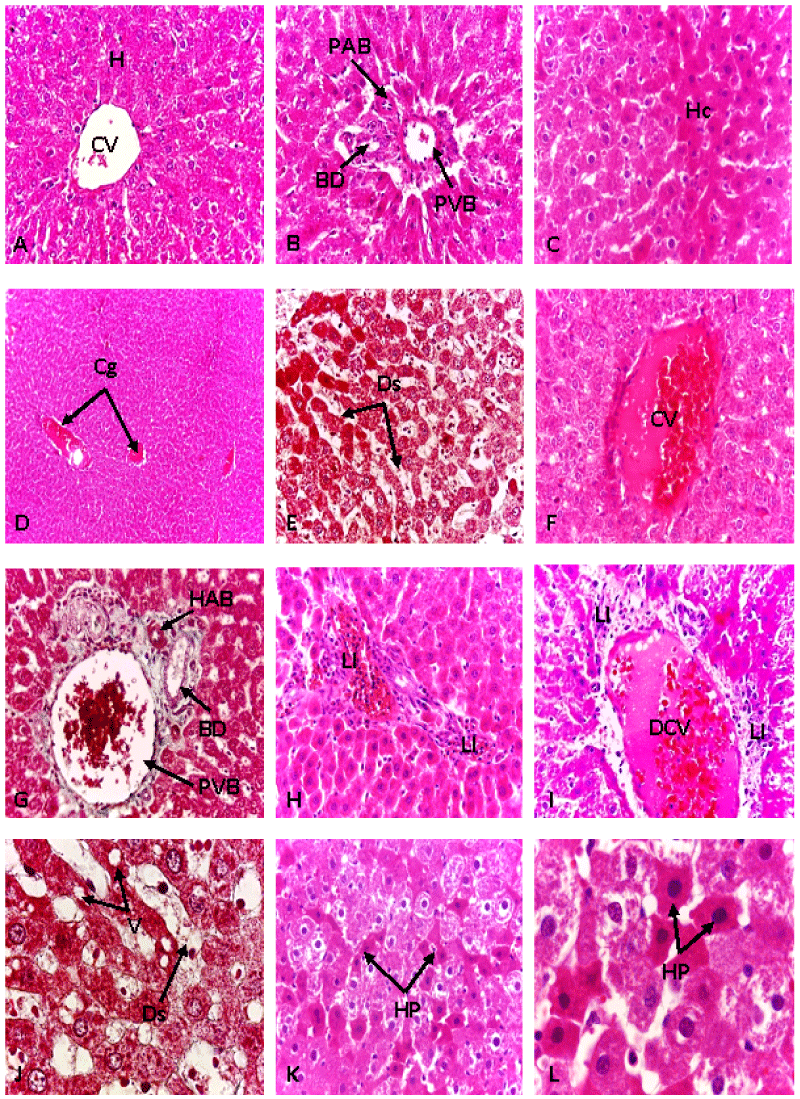
| Figure 3: Photomicrographs of rat liver control and treated. The control (A and B, H&E 400x) reveal normal liver parenchyma with central vein (CV) and portal triad (HAB: hepatic artery branch; PVB: portal vein branch; BD: bile duct). The rat treated showing change in the liver parenchyma (C, H&E 400x), vascular congestion (Cg) (D, H&E 100x), dilatation of sinusoid (Ds) (E, Trichrome 1000x), dilatation of CV (F, H&E 400x) and hyperthrophy of portal triad (G,Trichrome 400x). In the parenchyma liver lymphoid infiltration (LI) (H, H&E 400x) and dislocation of CV (DCV) are also seen (I, H&E 400x). Cytoplasmic vacuolation of some cells (V) with marked dilatation of sinusoid capillaries (J, Trichrome 1000x). The hepatocyte showing loss of their normal architecture (K, 400x; L, 1000x; H&E). HP: Hepatocyte’s proliferation, Hc: hepatic cords. ( H: hepatocyte; Ds: dilated sinusoids; CV: centrolobular vein; Cg: vascular congestion; V: vacuolization; LI: lymphoid infiltration; DCV: dislocation of the wall of the central vein) |