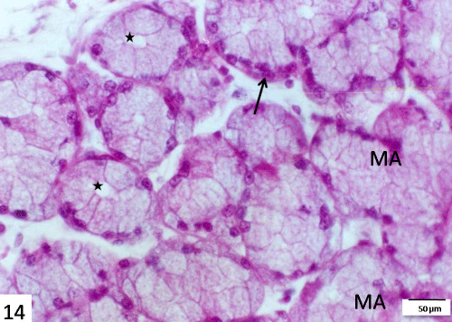
Figure 14:
A section in the submandibular gland of 14 days starved group stained with H&E showing distortion of mucous acini (MA), some of them with dilated lumens (star) and atrophied serous demilunes (arrow). Scale bar=50 μm.