|
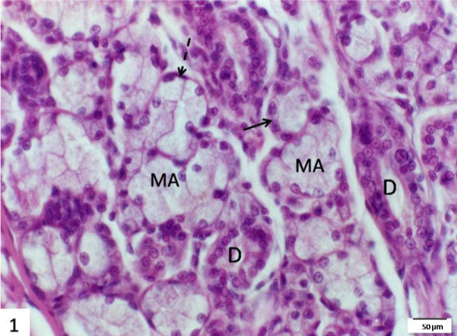
| Figure 1: A section in the submandibular gland of the control group stained with H&E showing mucous acini (MA) with cuboidal shaped cells and pale basophilic vacuolated cytoplasm as well as the serous demilunes (arrow). Note, the striated ducts (D) and the myoepithelial cells (dotted arrow) gasping the acini. Scale bar=50 μm. |