 |
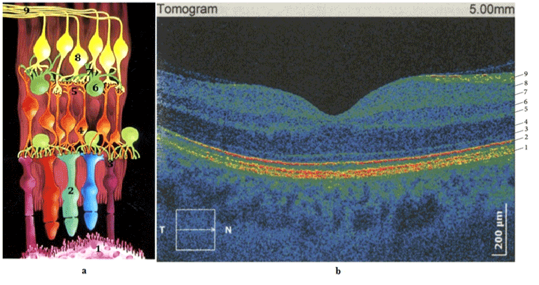
| Figure 1: Human retina (a) scheme of retinal morphology, (b) OCT scan, macular region (HD-SOCT Copernicus +), 1. Retinal pigment epithelium, 2. Inner and outer photoreceptor segments, 3. External limiting membrane, 4. Outer nuclear layer, 5. Outer plexiform layer, 6. Inner nuclear layer, 7. Inner plexiform layer, 8. Ganglion cell layer, 9. Neurofiber layer. |