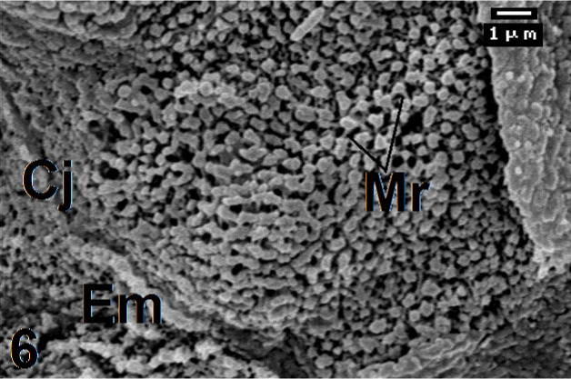
Figure 6:
Magnified micrograph from Figure 5 shows tiny microvilli, with enlarged apices (Em). Some are fused together and interconnected by microridges (Mr). Cj: Cell junction. ×7500.