|
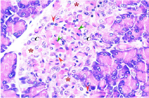
| Figure 3: A section from diabetic group 2 (G2.2) pancreas, showing loss of the cord arrangement of the cells with dilated congested capillaries (green heads) and lymphocytic infiltration (red arrows). It also shows necrotic cells having karyorrhectic nuclei and chromatin margination (C), other areas show loss of cells (stars) (H&E x1000). |