 |
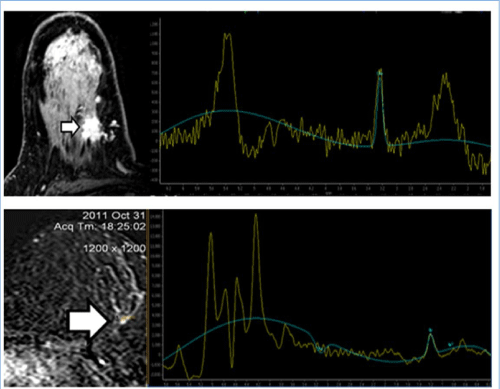
| Figure 5: (a) Left breast upper outer quadrant infiltrative mass lesion showing intense enhancement with evidence of high Choline peak in the MR Spectroscopy. (b) After complete NAC there is evidence of small residual enhancing focus with absence of Choline peak in the spectrum. |