 |
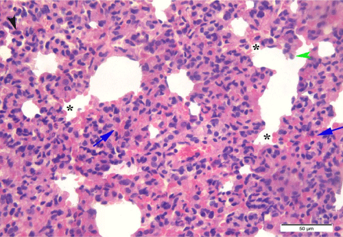
| Figure 7: A photomicrograph of a DEHP treated rat alveolar tissue showing marked thickening of interalveolar septa, interstitial hemorrhage (blue arrow), extravasated RBCs (*), necrotic type II pneumocytes with karyorrhectic nuclei (green arrow head). Some type II pneumocytes with mitotic figures are also shown (black arrow head). (H & E x400). |