 |
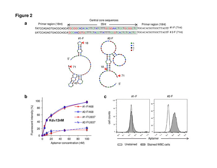
| Figure 2: Characterization of the developed aptamers. (a)Predicted two-dimensional structures of Aptamer sequences of class 1 (#1-F) and class 2 (#2-F) aptamers, including 5’primer, central core sequence, and 3’ primer regions, and predicted two-dimensional structures. (b) EpCAM-positive (MDA-MB-468) and -negative (U937) cells were exposed to Cy5-labeled #1-F and #2-F aptamers at indicated concentrations and the resultant cell binding fluorescence was quantified by flow cytometry. (c) White blood cells isolated from normal peripheral blood were incubated with #1-F and #2-F aptamers to rule out of target cell binding. All experiments were repeated by at least three times. |