 |
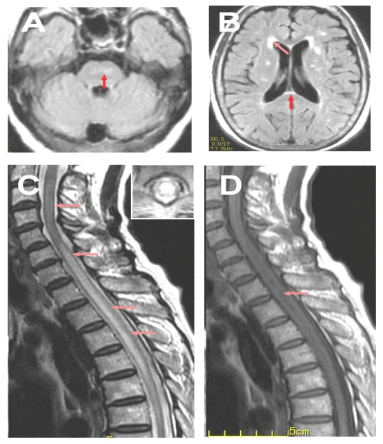
| Figure 1: MRI features of the case. A: Lesion in Pons (arrow); B: Brain lesions around the bilateral ventricle (arrow) and in corpus callosum (arrow); C: T2-weighed spinal MRI showed extensive lesion in the cervical and thoracic cord (C3-T5); D: T1-weighed spinal MRI showed hypointensity mainly around the central canal (arrow). |