 |
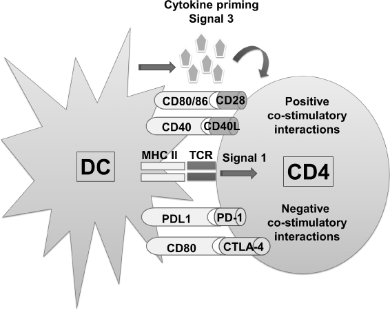
| Figure 1: Immunological synapse between a dendritic cell (DC) and a CD4 T cell. The scheme depicts the three signals between antigen presenting cells and T cells leading to T cell activation. Signal 1 is shown as binding between the peptide-MHC complex with the TCR, as shown in the center of the DC-T cell interaction. In the upper part, positive co-stimulatory interactions are shown, specifically CD80/CD28 and CD40/CD40L, while in the lower part of the DC-T cell interaction, negative co-stimulatory interactions are shown. In this case, PD-L1/PD-1 and CD90/CTLA-4. The integration within the T cell of these two types of interactions will determine the activation state of the T cell. On the upper part of the scheme, signal 3, or cytokine priming, is indicated. Depending on the combination of cytokines delivered by DC and T cells during their interaction, will result in different types of immune responses. |