 |
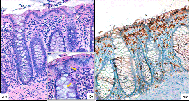
| Figure 5: Large bowel biopsy: H&E staining (on the left) shows a moderate lymphogranulocytic and plasmacellular inflammatory infiltration in the lamina propria. Intraepithelial lymphocytes show a basophilic nuclear chromatin pattern, irregular nuclear outline, and clear perinuclear halo (inset, yellow arrowheads). CD3 immunostaining (picture on the right) highlights the increase of the intraepithelial T lymphocytes (IELs), typical of lymphocytic colitis. Original magnification 200x; inset 400x. |