|
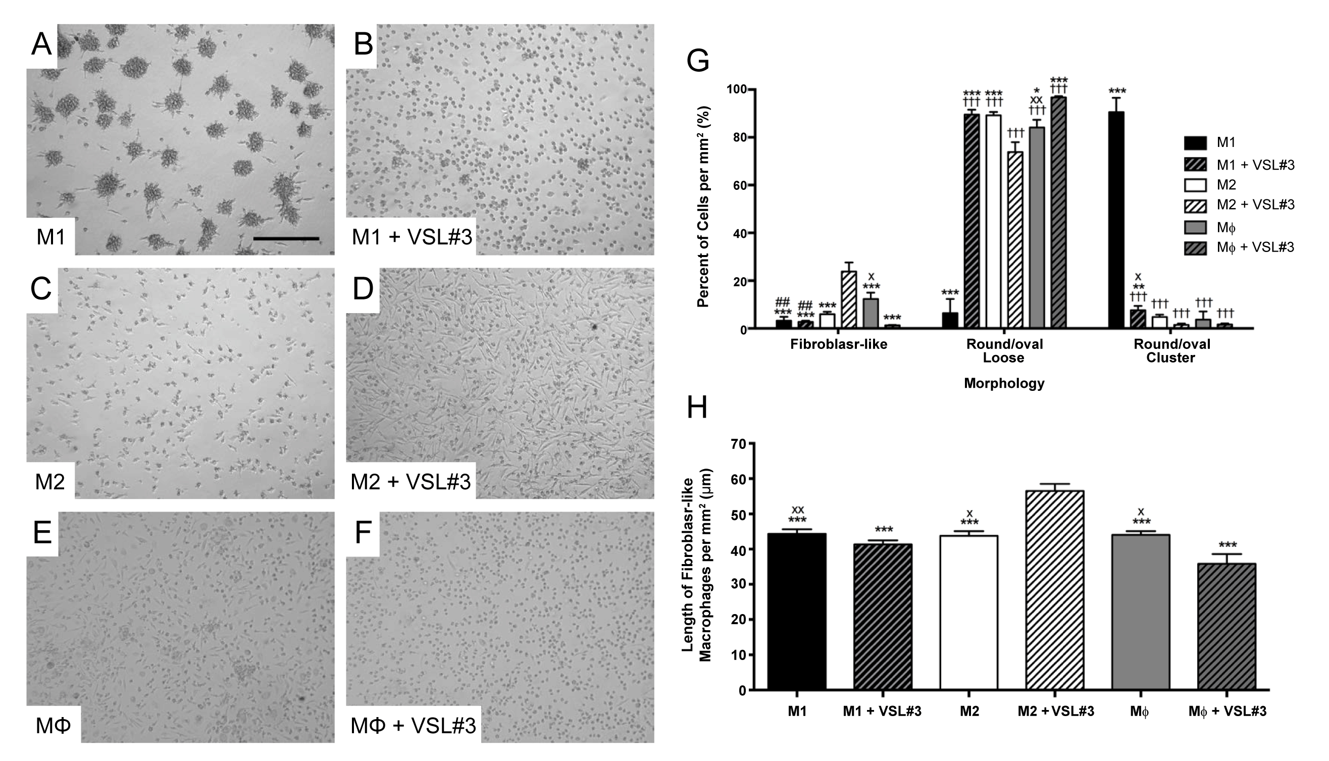
| Figure 2: VSL#3-induced changes in macrophage morphology. (A-D) Representative micrographs illustrating the morphology of untreated (A,C,E) and treated (B,D,F) M1, M2, and MΦ macrophages at day 11 of the protocol. Treatment consisted of exposure to 3.33x10⁷ CFU of the probiotic mixture VSL#3 for 3 days. Scale bar = 0.25 mm in A and applies to A-F. (G) Quantification of cells demonstrating different morphological characteristics. Cells were quantified by using the ‘cell counter’ and ‘analyze particles’ features of Image J. Two high-powered fields (mm2) were counted per well, one in the center of the well and another in the periphery. Data represents the mean ± SE of 10-12 counts per treatment, half in the center and half in the periphery. (H) Length of the fibroblast-like macrophages quantified in G, measured with ImageJ. Data were analyzed using one-way ANOVA and Tukey’s multiple comparisons test. *p<0.05, ***p<0.001 vs M2+VSL#3; ##p<0.01 vs MΦ; †††p<0.001 vs M1, xp<0.05, xxp<0.01 vs MΦ+VSL#3. |