 |
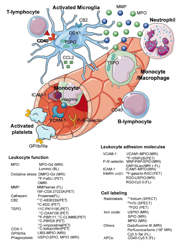
| Figure 1: Targets and probes in molecular imaging of neuroinflammation. Key cellular players in neuroinflammation are activated microglia, monocytes/macrophages, neutrophils, Tlymphocytes and B-lymphocytes. Microglia are resident brain leukocytes, and upregulate translocator protein (TSPO), cyclooxygenase 1 (COX1), and cannabinoid receptor 2 (CB2) under inflammatory conditions. Blood-borne leukocytes extravasate into the brain through interaction of cell surface integrins with specific endothelial adhesion molecules (e.g., ICAM-1, VCAM-1, P-/Eselectin). Once in the subendothelial space, exposure to chemokines (e.g. CCL2 released by microglia) further directs them towards their target. Stimulated cells then secrete effector molecules (e.g., matrix metalloproteinases [MMPs] and myeloperoxidase [MPO]), which trigger axonal damage and/or demyelination. Cell interaction between antigen-presenting cells (APCs, e.g., B-lymphocytes, microglia, dendritic cells) is mediated via CD40 amongst other molecules. Activated platelets can also produce reactive oxidative species and trigger thrombosis. BL: Bioluminescence imaging; FL: Fluorescence Imaging (Adapted from Servier Medical Art). |