 |
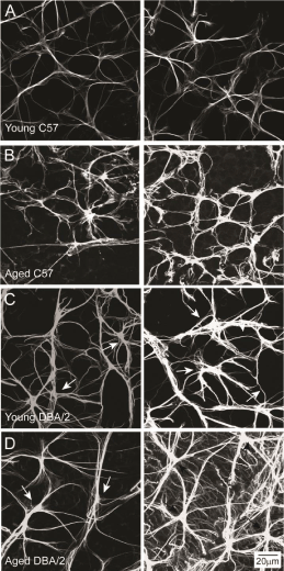
| Figure 1: Astrocyte morphology is variable across individual retina. Representative confocal micrographs of whole-mount retina from young C57 (A), aged C57 (B), young DBA/2 (C) and aged DBA/2 (D) mice immunolabeled with the astrocyte marker GFAP. Left and right columns illustrate variations in astrocyte morphology from comparable retinal areas within an individual retina. Glaucomatous retina (aged DBA/2) exhibits a broader range of astrocyte morphologies than young C57, aged C57 and young DBA/2 retina. Scale is the same for (A-D). |