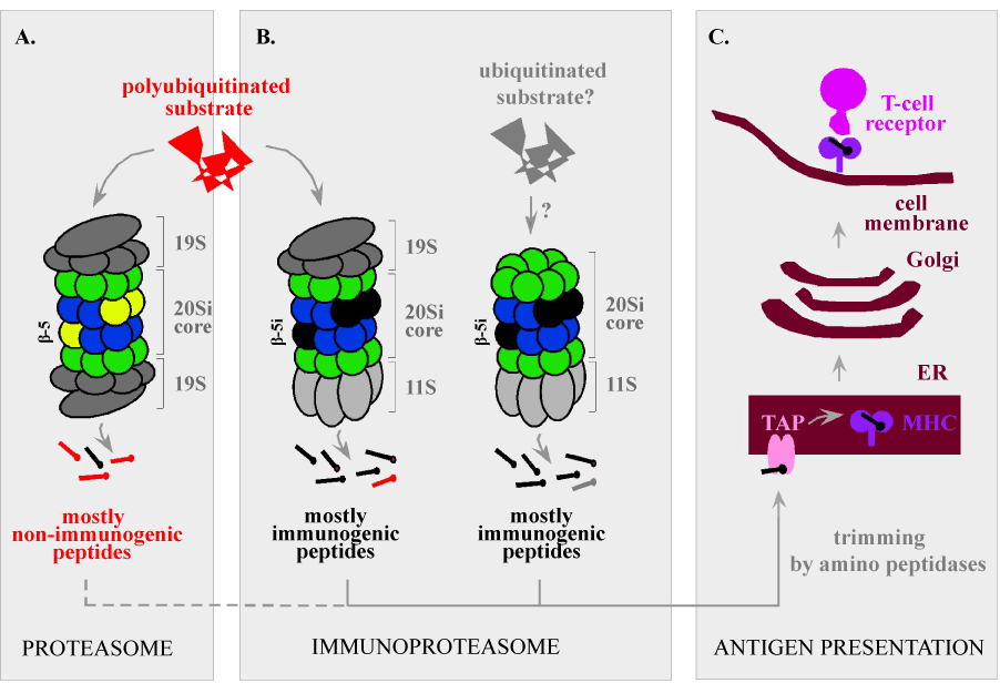
(A). In somatic cells not exposed to IFNs, the proteasome collaborates with the 19S activator and degrades polyubiquitinated substrates to peptides that are poor precursors for T-cell ligands.
(B). The immunoproteasome is more proficient in this role due to replacement of proteolytic subunits (yellow) with their immune versions (black) and collaboration with the 11S activator.
(C). Key steps leading to antigen presentation by MHC class I molecules. See text for details.