 |
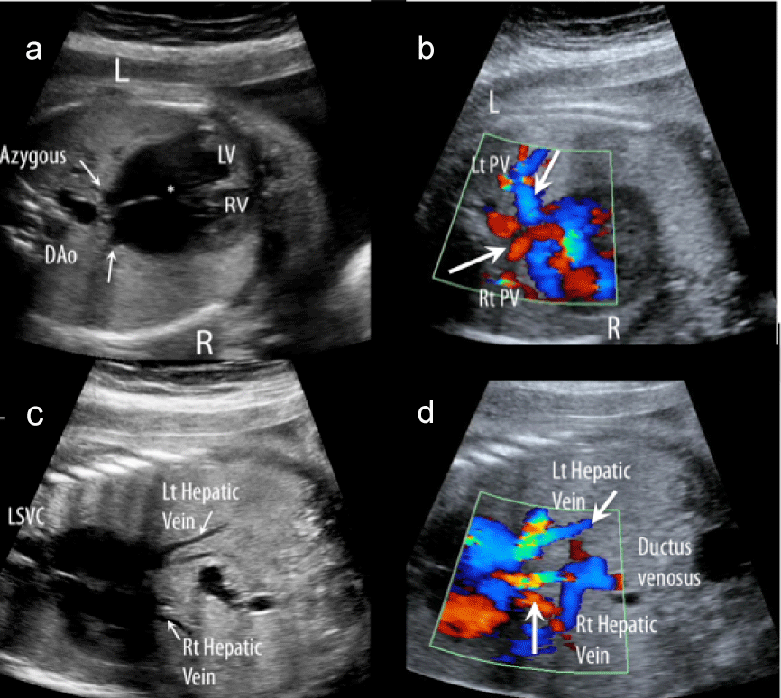
| Figure 1: Images from a 30 week fetus with left atrial isomerism, mesocardia and anomalous systemic and pulmonary venous connections. Left and right pulmonary veins (a & b, arrows) and left and right hepatic veins (c & d, arrows) can be seen connecting on either side of the atrial septum by 2D and color flow map imaging. A coronary sinoseptal defect was identified (*). The dilated azygous vein connected with a left superior vena cava that drained into the left sided atrium. Surgical repair with intra-atrial baffling of systemic and pulmonary veins was used to correct this lesion at 9 months. L-left, R-right, Lt-left, Rt-right, DAo-descending aorta, RV-right ventricle, LV-left ventricle, PV-pulmonary vein, LSVC-left superior vena cava. |