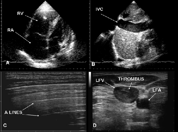
A: typically right chambers are anlarged, B: inferior vena cava is dilated, C: lung is “dry” and D: CUS shows a thrombus in the left femoral vein. RV= right ventricle, LV= left ventricle, IVC= inferior vena cava, LFV= left femoral vein, LFA= left femoral artery.