 |
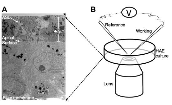
| Figure 3: (A) Transmission electron microscopy image of human airway epithelia (HAE) in vitro. Epithelial cells rest on a filter membrane and develop tight junctions towards the apical surface. A layer of airway surface liquid (ASL) 4 to 7 mm high lines the surface of airway epithelia. (B) Diagram of the system used for measurement of ASL glucose. HAE in a temperature-controlled chamber (not shown) are visualized with the lens of an inverted transmitted light microscope. The reference and working microelectrodes are mobilized with micromanipulators (not shown) until contact with the ASL is established. |