 |
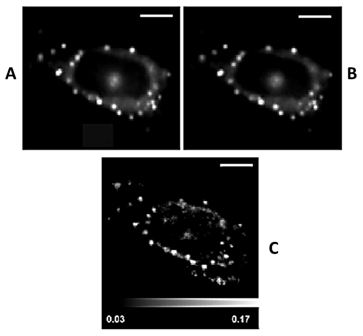
| Figure 5: Fluorescence intensity image of 1 with parallel (A) and perpendicular (B) polarizations relative to the incident light in HeLa cells as measured using an inverted microscope and polarizing image splitter. The fluorescence anisotropy image for 1 where bright spots have anisotropy values which correspond to a viscosity up to 70 cP. Reproduced by permission of ref. 24. |