 |
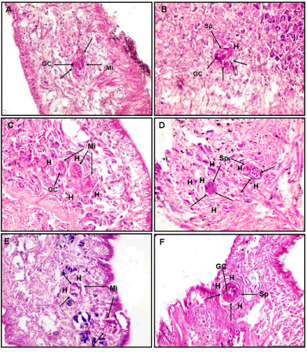
| Figure 4: T.S of Biomphalaria alexandrina snails exposed to Schistosoma mansoni miracidia (SBSC): left panel (6hr post exposure), right panel (72hr post exposure). A&B: Ismailia snails, A: miracidium Mi (arrows) in head-foot region; note successful penetration of miracidia without tissue response at this time, B: hemocytes H infiltration around the abnormally developed sporocyst Sp in the head-foot region as an inward step of forming a capsule, C&D: Kafr El- Sheikh snails, C: three encapsulated miracidia Mi (arrows) in head-foot region; note obvious tissue reactions through hemocytes H aggregations around the miracidia, D: hemocytes H infiltration around the abnormally developed sporocysts Sp in the head-foot region forming a capsule, E&F: Damietta snails, E: two miracidia Mi (arrows) in tentacle; Note haemocytic response H, F: degraded sporocyst Sp and large capsule formation by several layers of hemocytes H in the head-foot region near the tentacle; germinal cell (GC); tegument (T) (h & e) (x 400). |