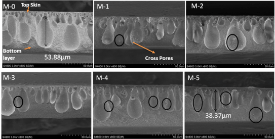
Figure 5:
The cross-section SEM images of the pure CAD (M-0) and the blend membranes (M-1, M-2, M-3, M-4 and M-5).