 |
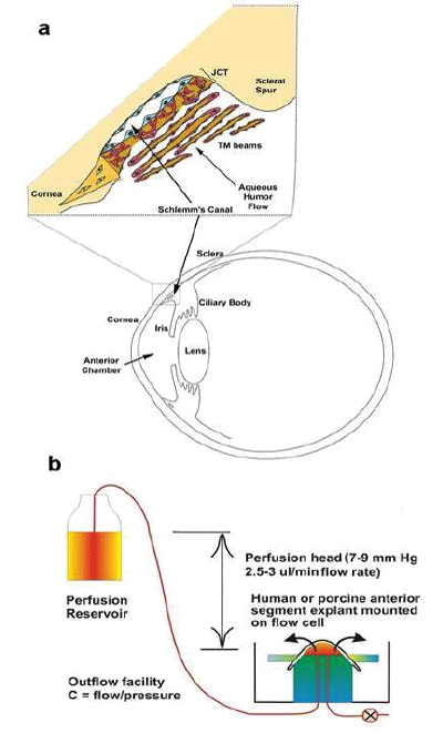
| Figure 1: Outflow pathway and perfused anterior segment organ culture model. a) Inset shows the human aqueous humor outflow pathway. Aqueous humor flows from the ciliary body through the pupil and out through the trabecular meshwork into Schlemm’s canal where it enters the venous drainage system. The TM outer beams are highly phagocytic. Aqueous humor percolates between the TM beams and then passes through the juxtacanalicular (JCT) region of the TM and across Schlemm’s canal inner wall endothelium [1-5]. Aqueous humor bathes the avascular cornea, lens and TM. Aqueous humor, formed by the ciliary body, flows at a relatively pressure-insensitive rate of around 2.75 μl/min. IOP is thus regulated by adjustments in the resistance to outflow that resides in the deepest portion of the JCT and Schlemm’s canal inner wall. (This diagram is modified significantly from an earlier review [1]. b) Anterior segments organ culture perfusion system [6] Anterior segments, including the cornea, TM and approximately 5mm of sclera but without the iris, ciliary body or lens, are clamped into a polycarbonate flow cell [6]. Culture media is perfused through ports in the bottom of the flow cell, driven by a defined pressure head selected to mimic the physiologic IOP minus the episcleral venous pressure, which is missing in the model. Fluid flow rates are measured gravimetrically. The system is maintained in a standard CO2 culture incubator at 37°C and 100% humidity [1]. |