 |
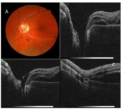
| Figure 6: Small, temporal inferiorly located congenital optic disc pit associated with macula detachment (case 2) and series of SOCT images a. optic disc pit with herniated dysplastic retina; b. posterior vitreous traction in the projection of optic disc pit, retinoschisis c. communication fistula between the pit and subretinal/ intraretinal space (white arrow). |