 |
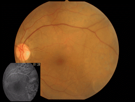
| Figure 4: Six months after initial presentation, fundus photograph of the left eye displays 2 almost stable choroidal tumors at superior and temporal area. Fluorescein angiography shows clearly 2 choroidal tumors with contrast medium leaking (shown as an inset). |