 |
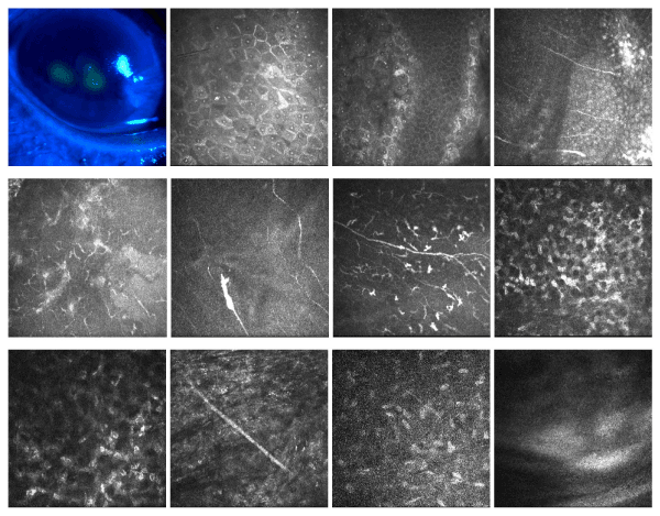
| Figure 1: HRTII-RCM images (300 Ám by 300 Ám). (A) The primary HSK cornea under the slit lamp microscope. (B) Epithelial cells loosing and oedema. (C) Normal epithelial cells and degenerating cells. (D,E) The broken subepithelial nerves in the lesion. (F) The broken nerve and expanding stump. (G) Quantity of DCs near the nerve. (H,I,J,K) The stromal cell oedema, no inflammatory cells or neovascularisation in the stroma, and stromal nerve thickening. (L) No endothelial cell damage. |