 |
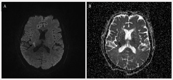
| Figure 1: MRI of the brain demonstrating acute infarct. (A) MRI demonstrates hyperintensity within the right internal capsule on diffusion weighted images (black arrow). (B) Acute infarction is confirmed by hypointensity within the same territory on apparent diffusion coefficient (white arrow). |