 |
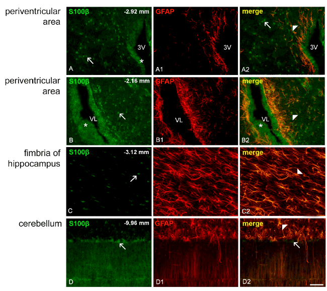
| Figure 3: Photomicrographs of brain coronal sections of the Wistar rat double labeled by immunofluorescence (GFAP/red and S100B/green). Arrowheads indicate the high S100B and GFAP colocalization observed in the periventricular areas (yellow in A2 and B2), in the fimbria of the hippocampus (yellow in C2) and in the cerebellum (yellow in D2). Arrow indicates S100B-IR cells. Note that many S100B-IR cells (green) are not also GFAP IR (red) in A2 and D2. Asterisks indicate S100B-IR ependymal cells. Scale bar: 100 μm. |