 |
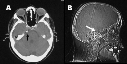
| Figure 1: Position of the electrode in the foramen ovale during percutaneous thermocoagulation. A: procedure performed under CT control; the black arrow indicates the position of the electrode in the foramen ovale at axial CT image. B: procedure performed under fluoroscopic control (lateral cranial radiographic image): the white arrow indicates the retrosellar position of the electrode, used to achieve selective anesthesia localized to the only III trigeminal division. |