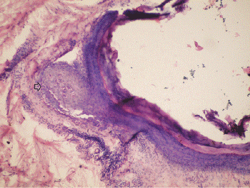
Figure 3:
Comedonal Darier’s disease: Histopathological findings from a nodular lesion. A markedly dilated hair follicle cyst with acantholysis and prominent dyskeratosis involving the wall of the cyst (Arrow) (H&E, original magnification × 40).