 |
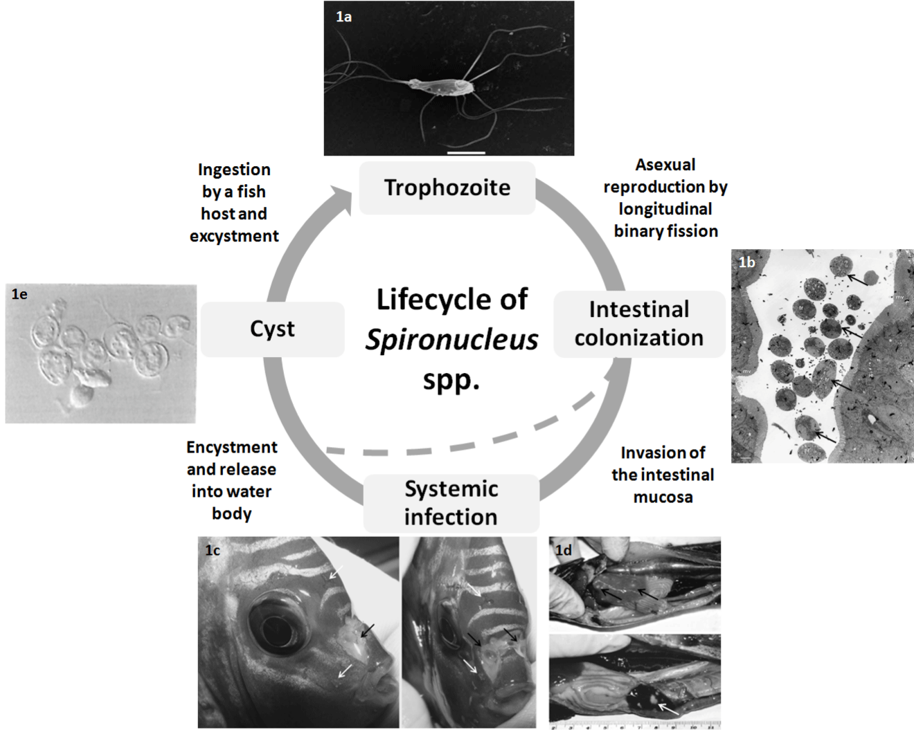
| Figure 1: Depiction of the typical life cycle of Spironucleus spp. Flagellated Spironucleus trophozoite resides in the intestinal tract of the fish host, scale bar = 5 μm (a). Multiplication of the parasite leads to intestinal colonization (b), arrows indicate presence of numerous trophozoites in the intestinal lumen (transverse section). Invasion of the intestinal mucosa leads to systemic infection, manifesting as hole-in-the-head disease in ornamental fish (c), white arrows indicate small, bilaterally symmetrical lesions, black arrows indicate larger lesions which have coalesced (pers. obs.). Granulomatous nodules (indicated by arrows) in the liver and spleen are associated with systemic Spironucleosis in Salmo salar (d). Transmission of the parasite through the water body is facilitated via an encysted form (e), following intestinal colonisation or systemic infection. Images adapted from Paull and Matthews [24] with permission from Inter-Research (a), Poynton et al. [46] with permission from Inter-Research (b), pers. obs (c) Guo and Woo [26] with permission from Inter-Research (d) and Uldal [51] with permission from The European Association of Fish Pathologists (e). |