 |
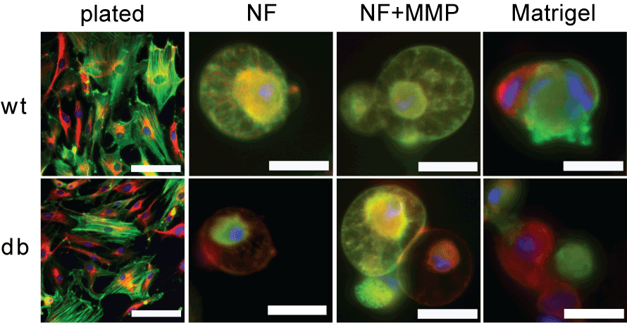
| Figure 3: Staining for fibroblast phenotype. Cells were stained with antibodies for fibroblast marker vimentin (red) and myofibroblast marker a-smooth muscle actin (a-SMA, green) and DAPI (blue). The top panels are wild type cells and the bottom panels are diabetic cells. Cells were either plated on gelatin coated dishes (left panels) or embedded in NF, NF+MMP, and Matrigel scaffolds (higher magnification panels, from left to right). All cells were positive for fibroblast marker vimentin and approximately 70-80% of cells were also positive for myofibroblast marker a-SMA. Scale bar in left panels is 100Ám. Scale bar in higher magnification panels is 25Ám. |