 |
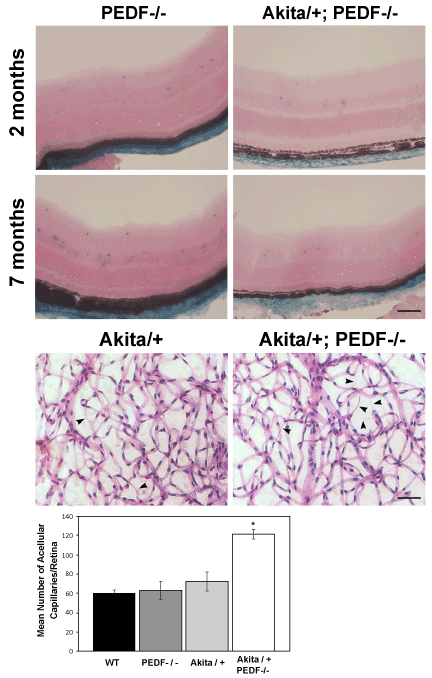
| Figure 7: Increased acellular capillaries in retinas from Akita/+; PEDF-/- male mice. Retinal sections prepared from 2 and 7 month old PEDF-/- and Akita/+; PEDF-/- male mice were stained for β-galactosidase activity (Panel A). Note β-falactosidase activity in the inner and outer plexiform layer of retinas from Akita/+; PEDF-/- male mice was decreased compared with PEDF-/- male mice. In Panel B, trypsin digestion of the retinal vasculature were prepared from 9 month Akita/+ and Akita/+; PEDF-/- male mice and acellular capillaries were quantified. Arrow heads point to acellular capillaries. Please note that more acellular capillaries formed in Akita/+; PEDF-/- male mice compared to Akita/+ male mice (*P < 0.05; scale bar= 50 μM; n ≥ 5 mice). |