|
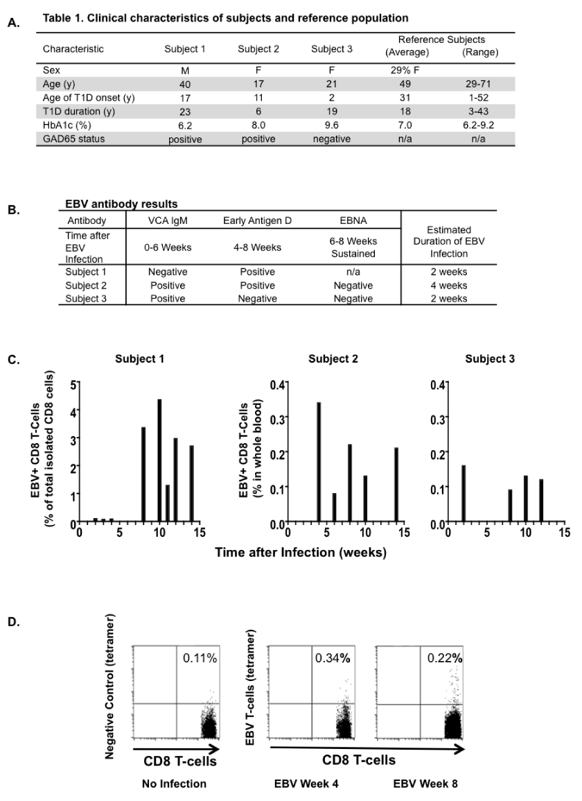
| Figure 1: Clinical characteristics and time course of EBV infection. A. Summary of clinical characteristics for the three EBV subjects. B. Results for EBV antibody testing on blood samples drawn during the first visit for the three EBV subjects and correlation with estimated start of EBV infection. C. Time course of the appearance of EBV-specific T-cells in Subject 1, Subject 2 and Subject 3. As detailed in the Materials and Methods, the staining for EBV reactive T-cells in Subject 1 was performed on isolated CD8 T-cells; the staining for EBV reactive T-cells for Subject 2 and Subject 3 was performed on whole blood lysates with CD8 T cells identified with CD8 antibody. In the graphs, time point 0 indicates the estimated occurrence of EBV infection, based on the timing of the appearance of antibodies directed against EBV. D. Representative dot plots of flow cytometry results. Level of non-specific tetramer binding by CD8 T-cells was determined using a tetramer loaded with an irrelevant peptide sequence (Negative Control, left panel). The middle and right panels show examples of positive staining using EBV-specific tetramers. |