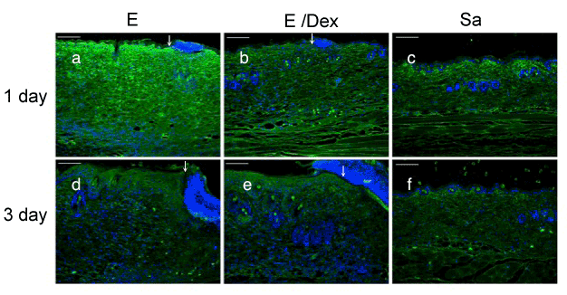
Sections of skin tissue sites after injection of E (A,D), E/Dex (B,E), Sa (C,F) were prepared on days 1 (A,B,C) and 3 (D,E,F) and stained using a biotinylated hyaluronic acid-binding protein. DAPI (blue) was used for counterstaining. Borders between infiltrated (left side) and necrotic (right side) regions are indicated by arrows. Bars: 100 μm.