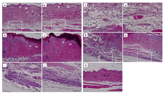
 (A) Dorsal skin sections, prepared on days 1 (A,B,C,D), 3 (E,F), and 5 (G,H,I,J,K) after injection of E (A,C,E,G,I), E/Dex (B.D,F,H,J), and Sa (K), were stained with HE.
Border areas between infiltrated regions (IRs; left side of lines) and necrotic regions (NRs; right side of lines) are indicated by dashed lines. Boxed areas of A and
B are enlarged in C and D. Boxed areas of G and H are enlarged in I and J. Arrows show polymorphonuclear leukocytes. Arrowheads show regeneration regions of
muscle fiber. Bars: A,B,E,F,G,H and K, 100 μm; C,D.I and J, 50 μm.