 |
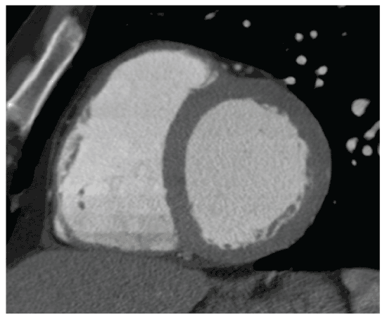
| Figure 1: End diastolic image of the left ventricle. Contrast enhancement of the EBCT image allows clear distinction of the endocardial border. Utilizing the computerized program in the scanner, each level is divided into 12 equal segments and compared to the end systolic image at the same level. The program then calculates an ejection fraction for each segment. |