 |
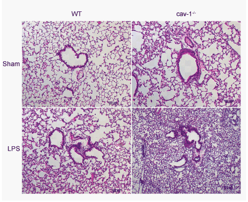
| Figure 2: Inflammatory infiltration into lung tissue following LPS treatment Following BAL, lungs were inflated with 4% formaldehyde by gravity. Paraffin embedded sections were cut and stained with H&E. Sham treated cav-1-/- mice (cav-1-/- sham) have a baseline pathology of increased cellularity (non-inflammatory) compared to WT (WT sham). Following LPS treatment, perivascular and peribronchiolar infiltrates are found in WT mice (WT LPS) and this inflammation is increased in cav-1-/- mice (cav-1-/- LPS). Representative images taken at 10X by light microscopy and are representative of sections from three mice per group. The scale bar is 100 mm in all photomicrographs. |