 |
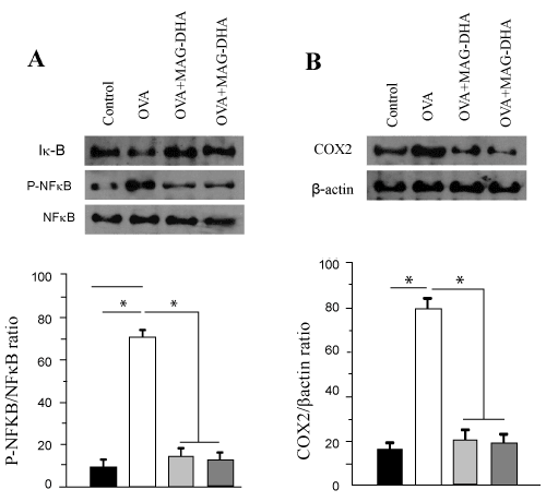
| Figure 3: Effect of MAG-DHA and MAG-EPA on airway inflammatory markers in lung tissue. A: Western blot and quantitative analysis of lung homogenate fractions derived from Control, OVA, OVA + MAG-DHA and OVA + MAG-EPA, using specific antibodies against IkBa, p65 NFkB and its phosphorylated form (n = 6). B. Western blot and quantitative analysis of COX2 protein detection. Staining densities in the homogenates were expressed as a function of b-actin signals (n=6). * P < 0.05. |