 |
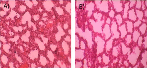
| Figure 5: A) Photomicrograph of section of fetal lung (control), H&E; 400X. B) Photomicrograph of section of fetal lung (experimental) showing evidence of atelectasis, interstitial oedema with focal areas of type-2 pneumoyte hyperplasia (black arrow heads), H&E; 400X. |