 |
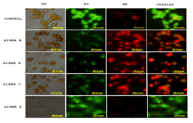
| Figure 5: Apoptosis is evidenced by AO/EB double staining. Morphological study of A549 cells cultured in 8 well Lab-Tek II chamber slide and treated with 400μM of various sequences of CCTα-siRNA: siRNA A, B, and C and siRNA S for 24 hr. The figure represents images viewed and captured by Fluorescence microscope coupled with Olympus camera. After incubation, cells were stained with AO/EB to detect apoptosis. Images were captured at different settings, BF: Bright Field; AO: Acridine Orange; EB: Ethidium Bromide. The last column represents the overlay images of AO and EB. Viable cells excluded ethidium bromide and their nuclei were bright green with intact structure, while apoptotic cells were orange to red color with highly condensed nuclei. |