 |
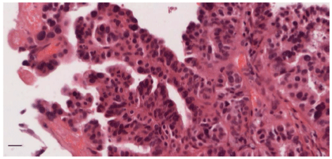
| Figure 1: Micropapillary features in an urothelial tumour from a patient with a history of CE.Micropapillary variant UC exhibits surface micropapillary features and small clusters of tumor cells that lack a fibrovascular core (x40 magnification). The patient had a cystoprostatectomy 10 weeks after initial transurethral resection of the bladder tumor. There was no residual tumor. The patient has recovered very well. (Scale bar 20 μm). |