Case Report Open Access
Are There Risk Factors for Splenic Rupture During Colonoscopy? Case Report and Literature Review
| Garancini Mattia1*, Maternini Matteo1, Romano Fabrizio1, Uggeri Fabio1, Dinelli Marco2 and Uggeri Franco1 | |
| 1Department of General Surgery, San Gerardo Hospital, University of Milano Bicocca, Monza (MI), Italy | |
| 2Department of Digestive Endoscopy, San Gerardo Hospital, University of Milano Bicocca, Monza (MI), Italy | |
| Corresponding Author : | Garancini Mattia, MD Department of General Surgery San Gerardo Hospital, University of Milano Bicocca Via Pergolesi 33, 20052, Monza (MI), Italy Tel: 039 233 3600 Fax: 039 233 3600 E-mail: mattia_garancini@yahoo.it |
| Received September 22, 2011; Accepted November 11, 2011; Published November 13, 2011 | |
| Citation: Garancini M, Maternini M, Romano F, Uggeri F, Dinelli M, et al. (2011) Are There Risk Factors for Splenic Rupture During Colonoscopy? Case Report and Literature Review. J Gastroint Dig Syst S2:001. doi: 10.4172/2161-069X.S2-001 | |
| Copyright: © 2011 Mattia G, et al. This is an open-access article distributed under the terms of the Creative Commons Attribution License, which permits unrestricted use, distribution, and reproduction in any medium, provided the original author and source are credited. | |
Visit for more related articles at Journal of Gastrointestinal & Digestive System
Abstract
Background: Splenic rupture is an uncommon but potentially fatal complication of colonoscopy.
Objectives: A case of splenic rupture during colonoscopy is reported and a review of literature is presented focusing the attention on evaluation of potential risk factors.
Case Report: We report the case of a 77 years old man who developed splenic rupture during colonoscopy diagnosed with CT scan and treated with splenectomy.
Results: More than 70 articles and more than 90 cases were found in the world literature; the review revealed that splenic rupture occurred more frequently in female, CT scan was the treatment was the referring diagnostic procedure in the large part of cases, splenectomywas the treatment of choice. On the other side none of the analyzed factor appeared as meaningful risk factors.
Conclusion: The knowledge of this complication is the best tool to aid in early diagnosis. Evaluation of hemodinamic status and CT scan play remarkable roles to resolve to the correct management and splenectomy remains the option chosen in the most part of cases.
| Keywords |
| Splenic injury; Splenic rupture; Trauma; Colonoscopy; Literature review |
| Introduction |
| Colonoscopy is an invaluable and largely used diagnostic and operative tool. It is considered a safe procedure with low complication rate. The most frequent complications are haemorrhage (with an incidence of 1-2%, usually associated with operative procedure like polipectomy) and colonic perforation (with an incidence of 0,1-0,2%) [1-4]. Other rare and unusual complications are pneumothorax, pneumomediastinum, appendicitis, small bowel perforation, septicemia, incarceration of hernia, pneumoscrotum, mesenteric tears, retroperitoneal abscess and colonic volvulus. |
| In this report a case of splenic injury occurred during a colonoscopy in a patient carrier of ileo-colic Crohn’s disease is described; we also reviewed the literature about this rare complication of colonoscopy with a focus on individuation and analysis of risk factors. |
| Case Report |
| A 77-year-old man with a previous segmental ileal resection for Crohn’s disease, in regular surveillance with 5-acetylsalycilate, underwent colonoscopy because of bowel disorder and increased erythrosedimentation rate and C-reactive protein levels. His medical history included myocardial infarction, arterious hypertension and uninvestigated dyspeptic symptoms empirically treated in the past with Proton Pump Inhibitor. The procedure was performed with standard sedation (meperidine 40mg + midazolam 2,5mg intravenous) and proceeded as far as the terminal ileum without any difficulty during the intubation of the colon. Endoscopic findings were active Crohn’s disease of the ileocecal valve and terminal ileum. The procedure was well-tolerated and the patient was discharged home after 1 hour recovery time. Eight hours later he presented to emergency room of our hospital complaining left abdominal pain with Kehr’s sign positive (pain radiating to the left shoulder tip), fatigue and sustained hypotension. At assessment his heart rate was 95 bpm and blood pressure was 75/55 mmHg. His haemoglobine levels had fallen from 12.9g/dL, as determined 2 weeks before to the procedure, to 10.4 g/dl; platelet count and coagulation setting were normal. The abdomen was soft and mild-distended with generalized tenderness and with abdominal pain localized in left quadrants and in mesogastrium; bowel sounds were reduced. |
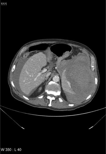
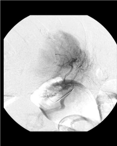
| The abdominal X-ray showed no free air and a nonspecific bowel gas pattern. No signs of rectal bleeding nor bleeding at gastric lavage were evident. Fluids replacement quickly improved the blood pressure and the clinical status, and surgeon decided to observe the evolution during the night. Eighteen hours after the endoscopic procedure a new hypotension episode (heart rate: 100 bpm and blood pressure: 70/50 mmHg) associated with persistent abdominal pain occurred and a Computerized Tomography scan was performed showing haemoperitoneum and a large splenic subcapsular haematoma (Figure 1). An urgent angiography was performed, but no demonstration of active bleeding was found (Figure 2). |
| Therefore the patient was transfused with 3 units of allogenic erythrocyte concentrates and surgeon planned laparotomy because of haemodynamic instability. A massive haemoperitoneum (with more than 1,5 litres of blood) and a large hematoma overlying the surface of the spleen with complete laceration of the splenic capsule were found. No peritoneal adhesion or anatomical abnormalities were discovered. Splenectomy was performed and pathological examination on the specimen revealed a parenchymal injury 6 cm long at the lower pole with no primitive disease of the spleen. After surgery the patient received standard post-splenectomy vaccinations (anti S. pneumoniae, H. influenza and N. meningitides) and was discharged home on the 7th post-operative day. |
| Methods |
| We performed a research on Pubmed-Medline entering as keywords “splenic rupture”, “splenic injury” and “splenic trauma” alone and in association with “colonoscopy”; in this review all the articles were analyzed in full text version. Information regarding age and gender of patients, type of endoscopic procedure (diagnostic, performance of biopsy or polipectomy), presence of risk factors (previous abdominal surgery, presence of inflammatory bowel diseases or other intestinal/ abdominal pathologies, aspirin or anticoaugulant intake, etc), onset of symptoms, clinical presentation at time of diagnosis of splenic rupture, diagnostic modalities and treatment (splenectomy, conservative, other therapies) were collected and analyzed. |
| A special attention was ascribed to individuation of risk factors. In particular for previous abdominal surgery was considered every operative surgical abdominal procedure, with exclusion of minimally invasive diagnostic procedure like diagnostic laparoscopy for infertility [5]. |
| Results |
| In the present research more than 70 articles [5-81] and more than 90 cases were found in the world literature (Table 1 data included as supplementary). |
| In our review mean age was 63 years (range 29-90) and gender was male in 35/88 (39,7%) cases and female in 53/88 (60,3%) cases (9 with gender not reported). Onset of symptoms occurred within 24 hours after the procedures in 76/94 (80,8%) of the patients, while the remnant 18/94 (19,2%) of the patients had a delayed presentation. |
| up to several days (range: less than 1 hour to 12 days). There was no correlation between delayed presentation and conservative management of the complication, and probably onset of symptoms occurred 5-6 days after the procedure was related to rupture of a subcapsular haematoma. |
| The most frequent presentations were severe abdominal pain (usually on the left flank, present in 88/93 reports, 94,6%), back pain, increasing adynamia, tiredness, collapse, vomit. Clinical evaluation revealed abdominal distension, tenderness to palpation in left quadrants of the abdomen, rare or no bowel sounds, Kehr’s sign positive, hypotension, high pulse rate, shock. Blood examinations were unspecific showing generic signs of bleeding, and gastro-intestinal perforation or intra-luminal bleeding must firstly be excluded with RX of the abdomen and digital rectal exploration. |
| Usually the abdominal pain was the first symptom and was followed by hypotension. So the physician should be suspicious in case of left lateral abdominal pain after colonoscopy, even if it usually occurs also in cases not complicated. If the pain is associated with hypotension or decreasing of hematocrit and haemoglobin rate and intestinal bleeding or perforation are excluded, a study the abdomen with ultrasound and/or Computered Tomography (CT scan) should be considered mandatory. |
| Computerized Tomography scan is considered the referring diagnostic procedure for splenic trauma by the American Association for the Surgery of Trauma Organ Injury Scale [82]; in this review in 72/96 patients (75%) diagnosis was obtained with a CT scan. If the patient remains haemodynamically unstable, urgent explorative laparotomy is the only suitable management. Our review shows that in 18 on 96 patients (18,7%) diagnosis was demonstrated with laparotomy without any other diagnostic tool for an instable hemodynamic condition; most frequently these patients are referred in articles published before 1993, but even in recent years in some cases the diagnosis was intra-operative. The use of paracentesis to demonstrate hemoperitoneum [10] or the use of angiography as the only diagnostic tool to demonstrate active bleeding [6] has been abandoned in the 2 last decades, even if angiography conserved even in recent years a successful therapeutic role in case of demonstration of active bleeding with CT scan. |
| Ultrasound was often the first radiological step, but is usually followed by a CT scan for a definitive diagnosis; in this review only Ong et al. [22] in 1991 and Shah et al. [41] in 2005 used ultrasound as the only radiological tool (2/96, 2%) and in both of them after ultrasound a splenectomy was performed. |
| Both operative and non-operative treatment have been applied to patients with splenic rupture after colonoscopy. Conservative management should include broad spectrum antibiotics, intravenous fluids, blood transfusions (if necessary) and hemodynamic monitoring. In our review splenectomy was the most frequent treatment and was performed in 72/97 patients (74,2%), a conservative treatment without any invasive procedure was the choice option in 20/97 patients (20,6%), successful splenic artery embolization was performed in 4/97 patients (4,2%) [35,57,64,72], 1/97 patient was treated with laparotomy and wrapping the spleen in a Vicryl net [60]. One patient had a postmortem diagnosis, so the therapeutic options were not evaluated on a certain diagnosis as like the other patients and he was excluded from the “conservative management” group [21]. Mortality was reported in 2/97 cases (2%) [10,21]. |
| Evaluation of hemodynamic status and of CT scan of the abdomen are the priorities to determine the therapeutic option and represent the factors those predict failure of non- perative management; in this sense contrast enhanced CT scan is considered a key component of nonoperative treatment [83,84] even if in specialized hospitals real-time contrast-enhanced ultrasonography is already playing and important role in evaluation of active abdominal bleeding [85]. |
| Rao et al. report a review of 9 cases of splenic rupture after colonoscopy and recognized 5 associated factors those may play a role in splenic injury: rapid completing time, chronic history of smoking, propofol sedation, inadeguate colon clean-out, daily aspirin intake [86]. Risk factors reported by Rao at al. [86] are not evaluated in our review for lacking of these information in almost the totality of cases reported; on contrary Rao et al. [86] did not reported specific information of the cases reviewed, so their patients were excluded from our review. |
| Discussion |
| Splenic injury during colonoscopy was described for the first time in 1974 by Wherry and Zehner [5]. It’s not so easy to calculate the real incidence of this complication, and underreporting is probably one of the most important reasons. In our experience the first case occurred after 79000 procedures. Some groups in literature reported higher incidence of 1 in 6000-7000 colonoscopies [3,22,30,87] but some other authors reported no splenic injuries in large series respectively of 13580 and 30463 procedures [4,88]. Kamath et al. [73] reported 4 cases in 296000 colonoscopies (incidence: 0,001%). Splenic injuries clinically evident occurred during colonoscopy are really rare, even if the incidence of minor splenic injuries clinically not detectable is probably higher. The etiology of splenic injury during colonoscopy is related to a mechanical trauma occurred during the procedure; the consequence of this trauma is the partial or total avulsion of the splenic capsule and/or parenchymal laceration or fracture [27], subcapsular hemorrhage [29,30,32,40,51], rarely bleeding from splenic vessels at the hilum [58,74]. |
| The precise mechanism is still not yet clarified. Many authors indicate as causes of this trauma excessive traction of the spleno-colic ligament in presence or not of short spleno-colic ligament or other causes of reduced mobility between the colon and the spleen like adhesion between spleen and splenic flexure, capsular thickening and fibrosis. Direct trauma to the spleen during colonoscopy has also been recognised as the cause of splenic rupture [26]. |
| It is interesting to know that also another endoscopical procedure like Endoscopic Retrograde Colangiopancreatography (ERCP) has splenic rupture as a possible rare complication [89,90]. Even for splenic rupture during ERCP an excessive traction of splenic’s ligaments is supposed to have a key role. On the other side a research conducted on Pubmed-Medline revealed that no case of splenic injury as a complication of gastroscopy is reported in literature. Gastroscopy is a procedure that usually is less hard-working than ERCP and can probably cause less important traction on the splenogastric ligament. The spleen is a relatively frail organ and probably the risk of splenic rupture during invasive endoscopic procedures that may cause traction on splenic ligaments is higher if the procedure is hard working. |
| In this review the authors individuate 3 classes of risk factors: primitive splenic pathologies, abdominal alterations and intestinal diseases and mechanisms procedure related or operator-related. It is not possible to calculate the real role of risk factors because this complication is really unusual. Our purpose is to evaluate the supposed and theoretical risk factors reported by many authors and our method is to analyze remote anamnesis, case history, type of endoscopical procedure (operative or not), intraoperative and anatomopathological findings. Unfortunately some case reports are very poor of informations and lack in some of these data; we calculated percentages on the number of articles with complete information. |
| Primitive splenic pathologies |
| Many authors suggest primitive splenic pathologies as possible risk factors. In this review one case of anatomopathological finding of splenic amiloidosis [49] and one case of small and medium-size vessels hyaline arteriosclerosis, compatible with longstanding hypertension [45] are reported. It is unclear the possible correlation of these anatomopathological finding with splenic rupture and no other cases of primitive splenic pathologies and in particular no case of splenomegaly in our review are known. |
| Abdominal alterations and intestinal diseases |
| Many authors indicate as risk factors: Crohn disease (that could be correlated with rigidity of the colon), multiple previous colonoscopies, peritoneal adhesion caused by previous abdominal surgery, previous pancreatitis, diverticulitis or other pathologies and tortuous left colon. In our review we found that 38/75 patients (50,6%) had previous abdominal surgery, 4/75 (5,2%) of patients have left tortuous colon (but probably presence of tortuous colon is sometimes unreported), 2 case of chronic pancreatitis whose 1 associated to pancreatic neoplasm [23,79], 1 case of endometriosis [58], 1 case of ulcerative colitis [65] and only 1 case prior our case reported presence of Crohn disease [7]. Moreover in our case Crohn disease was not correlated to presence of peritoneal adhesion and Crohn was ileal located; patients carriers of inflammatory bowel diseases (IBD) are usually under endoscopic surveillance and presence of just 3 cases in 75 patients (4%, 2 carriers of Crohn’s disease and 1 carrier of ulcerative colitis) indicates IBD as a not significant risk factor. Although presence of previous abdominal surgery in more than 50% of patients could appear meaningful, it probably has an inconclusive role as risk factor for two reasons. First, abdominal surgery doesn’t lead always to formation of adhesions and certain presence of peritoneal adhesions is rarely reported [7,12,13,42,50,71]. Second, in articles previously published evaluating predictive factors for difficult colonoscopies in series of 693 and 426 consecutive patients undergone to colonoscopies, a rate of respectively 49% and 35,2% patients with previous abdominal surgery, a percentage not so dissimilar from the one reported in this review [91,92]. |
| Mechanisms operation or operator-related |
| Operative colonoscopy, excessive traction on the splenic flexure during the procedure (like during the “hooking” of the splenic flexure to straighten the left colon, the “slide by” to go beyond the splenic flexure or the “alpha maneuver”), external application of abdominal pressure in particular on the left upper quadrant, supine position (some authors think that left lateral position should be preferred and that supine position increase the chance of splenic capsular tearing [33] are reported by many authors as possible risk factors. A few authors defined “difficult” some endoscopic procedures: for the unspecificity of the definition these data were not recordered, even if cases of laceration of splenic vessels at the hilum [58,74] or association of splenic rupture with colonic perforation probably confirms that hard-working procedure have a higher risk. Operative colonoscopy rate in our review was 28/95 (29,5%) (all of them submitted to polipectomy), and 9/95 (9,5%) of patients was submitted to biopsy; these data do not seem to be meaningful. Information about specific technical aspects of the endoscopic procedure are not reported in the major part of the reports. Some authors reported multiple previous colonoscopies as a risk factor, but the correlation with splenic injuries is not clarified. |
| Eight in 75 patients (10,6%) were under antiaggregant or anticoagulant therapies, and these are obviously risk factors for haemorrhage and theoretically could transform a subclinical microinjury in a clinically manifest active bleeding, even if in this review all the patients on medication with these kind drugs regularly stopped to take them some days prior the colonoscopy. Extremes of age which is supposed to be significant by many authors, in our opinion don’t have a predictive purpose. |
| Conclusion |
| Splenic rupture is uncommon but potentially fatal complication of colonoscopy, and we believe that this rare complication is actually not so rare. |
| The analysis of the literature shows that there are no major risk factors useful to predict splenic injuries during colonoscopy. There is no important correlation with IBD, primitive splenic pathologies, left tortuous colon or previous surgery. It’s very hard to understand the role of mechanisms operation or operator-related, in particular for the lack of information. In conclusion it’s clear that the knowledge of this complication is the best tool to aid in early diagnosis. Evaluation of hemodynamic status and CT scan play remarkable roles to resolve to the correct management and splenectomy remains the option chosen in the most part of cases. Colonoscopy is the optimal choice for colon cancer screening and is currently recommended by multiple medical societies, including the American Cancer Society, American College of Gastroenterology, and American Society of Gastrointestinal Endoscopy for patients≥50 years. It is still controversial whether splenic trauma should be mentioned on the consent form as a complication of colonoscopy, but the magnitude and severity of risks associated with colonoscopy are of paramount importance, given the otherwise healthy nature of the population undergoing screening. |
| Acknowledgements |
| Substantive contributions to the study was given by every authors in terms of data collection (Mattia Garancini, Matteo Maternini, Fabio Uggeri), editing of the case report (Mattia Garancini), editing of the review (Mattia Garancini, Fabrizio Romano), proof-reading (Franco Uggeri, Marco Dinelli). No financial support was necessary for this study. |
References
- Macrae FA, Tan KJ, Williams CB (1983) Towards safer colonscopy: a report on the complications of 5000 diagnostic and therapeutic colonscopies. Gut 24: 376-383.
- Schwesinger WH, Levine BA, Ramos R (1979) Complication in colonscopy. Surg Gynecol Obstet 148: 270-281.
- Smith LE (1976) Fiberoptic colonscopy and complications of colonscopy and polypectomy. Dis Colon Rectum 19: 407-412.
- Wexner SD, Garbus JE, Singh JJ (2001) A perspective analysis 13580 colonscopies: reevalutation of credentialing guidelines. Surg Endosc 15: 251-261.
- Goiten D, Goiten O, Pikarski A (2004) Splenic rupture after colonscopy. Isr Med Assoc J 6: 61-62.
- Telmos AJ, Mittal VK (1977) Splenic rupture following colonscopy. JAMA 237: 2718.
- Ellis WR, Harrison JM, Williams RS (1979) Rupture of spleen at colonscopy. Br Med J 1: 307-308.
- Kloer H, Schmidt-Wilcke HA, Schulz U (1984) Splenic rupture as a consequence of colonscopy. Dtsch Med Wochenschr 109: 1782-1783.
- Castelli M (1986) Splenic rupture: an unusual late complication of colonscopy. CMAJ 134: 916-917.
- Reynolds FS, Moss LK, Majeski JA, Lamar C Jr (1986) Splenic rupture following colonscopy. Gastrintest Endosc 32: 307-308.
- Doctor NM, Monteleone F, Zarmakoupis C, Khalife M (1987) Splenic injury as a complication of colonoscopy and polipectomy. Report of a case and review of the literature. Dis Colon Rectum 30: 967-968.
- Tuso P, McElligot J, Marignani P (1987) Splenic rupture at colonscopy. J Clin Gastroenterol 9: 559-562.
- Levine E, Wetzel LH (1987) Splenic trauma during colonscopy. Am J Roentgenol 149: 939-940.
- Walshe JJ, Lee JB, Gerbasi JR (1987) Continuous ambulatory peritoneal dialysis complicated by massive hemoperitoneum after colonoscopy. Gastrointest Endosc 33: 468-469.
- Lerone E, Wetzel LH (1987) Splenic trauma during colonoscopy. Am J Roetgenol 149: 939-940.
- Gores PF, Simso LA (1989) Splenic injury during colonoscopy. Arch Surg 124: 1342.
- Bier JY, Ferzli G, Tremolieres F, Gerbal JL (1989) Splenic rupture caused by colonoscopy. Gastroenterol Clin Biol 13: 224-225.
- Taylor FC, Frankl HD, Riemer KD (1989) Late presentation of splenic trauma after routine colonscopy. Am J Gastroenterol 84: 442-443.
- Merchant AA, Cheng EH (1990) Delayed splenic rupture after colonscopy. Am J Gastroenterol 85: 906-907.
- Rockey DC, Weber JR, Wright TL, Wall SD (1990) Splenic injury following colonscopy. Gastroentrol Endosc 36: 306-309.
- Colarian J, Alousi M, Calzada R (1991) Splenic trauma during colonscopy. Endosc 123: 48-49.
- Ong E, Bohmler U, Wurbs D (1991) Splenic injury as a complication of endoscopy: two case reports and a literature review. Endosc 23: 302-304.
- Viamonte M, Wulkan M, Irani H (1992) Splenic trauma as a complication of colonscopy. Surg Laparosc Endosc 2: 154-157.
- Dodds LJ, Hensman C (1993) Splenic trauma following colonoscopy. Aust N Z J Surg 63: 905-906.
- Heath B, Rogers A, Taylor A, Lavergne J (1994) Splenic rupture: unusual complication of colonscopy. Am J Gastroenterol 89: 449-450.
- Ahmed A, Eller PM, Schiffman FJ (1997) Splenic ropture: an unusual complication of colonscopy. Am J Gastroenterol 92: 1201-1204.
- Arnaud JP, Bergamaschi R, Casa C, Boyer J (1993) Splenic rupture: an unusual complication of colonscopy. Colo-proctology 6: 356-357.
- Coughlin F, Aanning H (1997) Delayed presentation of splenic trauma following colonscopy. SDJ Med 50: 325-326.
- Espinal EA, Hoak T, Porter JA, Slezak FA (1997) Splenic rupture from colonscopy: a report of two cases and review of literature. Surg Endosc 11: 71-73.
- Moses RE, Leskowitz SC (1997) Splenic rupture after colonscopy. J Clin Gastroenterol 24: 257-258.
- Reissmann P, Durst AL (1998) Splenic hematoma. A rare complication of colonscopy. Surg Endosc 12: 154-155.
- Olshaker JS, Deckleman C (1999) Delayed presentation of splenic rupture after colonscopy. J Emerg Med 17: 455-457.
- Tse CC, Chung KM, Hwang JS (1999) Splenic injury following colonscopy. Hong Kong Med J 5: 202-203.
- Melsom DS, Cawthorn SJ (1999) Splenic injury following routine colonscopy. Hosp Med 60: 65.
- Stein DF, Myaing M, Guillaume C (2002) Splenic ropture after colonscopy treated by splenic artery embolization. Gastrointest Endosc 55: 946-948.
- Rinzivillo C, Minutolo V, Gagliano G, Minatolo G, Morello A et al. (2003) Splenic trauma following colonoscopy. Giornale di Chirurgia 24: 309-311.
- Boghossian T, Carter JW (2004) Early presentation of splenic injury after colonscopy. Can J Surg 47: 148.
- Hamzi L, Soyer P, Boudlaf M, Najmef N, Abitbol M et al. (2003) Rupture splenique apre’s colonscopie : a propos d’un cas inhabituel sur venant sur une rate initialmente saine. J Radiol 84: 320-322.
- Prowda JC, Trevisan SG, Lev-Toaff AS (2005) Splenic injury after colonscopy: conservative managment using CT. AJR Am J Roentgenol 185: 708-710.
- Jaboury I (2004) Splenic rupture after colonscopy. Intern Med J 34: 652-653.
- Shah PR, Raman S, Haray PN (2005) Splenic rupture following colonscopy: rare in the UK? Surgeon 3: 293-295.
- Al Alawi I, Gourlay R (2004) Rare complication of colonoscopy. ANZ J Surg 74: 605-606.
- Lekas BJ (2004) Splenic hematoma as a complication of colonoscopy. J Am Geriatr Soc 52: 320-321.
- Wherry DC, Zehner H Jr (1974) Colonscopy-fiberoptic endoscopy approach to the colon and polypectomy. Med Ann Dist Columbia 43: 189-192.
- Naini MA, Masoompour SM (2005) Splenic rupture as a complication of colonoscopy. Indian J Gastroenterol 24: 264-265.
- Weisgerber K, Lutz MP (2005) Splenic rupture after colonscopy. Clin Gastroenterol Hepatol 3: A24.
- Zenooz NA, Win T (2006) Splenic rupture after diagnostic colonoscopy: a case report. Emerg Radiol 12: 272-273.
- Volchok J, Cohn M (2006) Rare complication following colonscopy: case reports of splenic rupture and appendicitis. JSLS 10: 114-116.
- Zerbi S, Crippa S, Di Bella C, Nobili P, Bonforte G et al. (2006) Splenic rupture following colonscopy in a hemodialysis patient. Int J Artif Organs 29: 335-336.
- Luebke T, Baldus SE, Holscher AH, Monig SP (2006) Splenic rupture: an unusual complication of colonoscopy: case report and review of the literature. Surg Laparosc Endosc Percutan Tech 16: 351-354.
- Shatz DV, Rivas LA, Doherty JC (2006) Management options of colonoscopic splenic injury. JSLS 10: 239-243.
- Johnson C, Mader M, Edwards DM, Vesy T (2006) Splenic rupture following colonoscopy: two cases with CT findings. Emer Radiol 13: 47-49.
- Pfefferkorn U, Hamel CT, Viehl CT, Marty WR, Oertly D (2007) Haemorragic shock caused by splenic rupture following routine colonscopy. Int J Colorectal Dis 22: 559-560.
- Lalor PF, Mann BD (2007) Splenic rupture after colonoscopy. JSLS 11: 151-156.
- Di Lecce F, Viganò P, Pilati S, Mantovani N, Togliani T, Pulica C (2007) Splenic rupture after colonoscopy. A case report and review of the literature. Chir Ital 59: 755-757.
- Tsoraides SS, Gupta SK, Estes NC (2007) Splenic rupture after colonoscopy: case report and literature review. J Trauma 62: 255-257.
- Holubar S, Dwivedi A, Eisendorfer J, Levine R, Strauss R (2007) Splenic rupture: an unusual complication of colonoscopy. Am Surg 73: 393-396.
- Janes SE, Cowan IA, Dijkstra B (2005) A life threatening complication after colonscopy. BMJ 330: 889-890.
- Cappellani A, Di Vita M, Zanghi A, Cavallaro A, Alfano G et al. (2008) splenic rupture after colonoscopy. Report of a case and review of literature. World J Emerg Surg 9: 3-8.
- Schilling D, Kirr H, Mairhofer C, Rumstadt B (2008) Splenic rupture after colonoscopy. Dtsch Med Wochenschr 133: 833-835.
- Famularo G, Minisola G, De Simone C (2008) Rupture of the spleen after colonoscopy. A lifethreatening complication. Am J Emer Med 26: 834.
- Guerra JF, San Francisco I, Pimentel F, Ibanez L (2008) Splenic rupture following colonoscopy. World J Gastroenterol 7: 6410-6412.
- Duarte CG (2008) Splenic rupture after colonscopy. Am J Emer Med 26: 117e1-3.
- Parker WT, Edwards MA, Bittner JG 4th, Mellinger JD (2008) Splenic hemorrhage: an unexpected complication after colonoscopy. Am Surg 74: 450-452.
- Saad A, Rex DK (2008) Colonoscopy-induced splenic injury: report of 3 cases and literature review. Dig Dis Sci 53: 892-898.
- Lewis SR, Ohio D, Rowley G (2009) Splenic injury as a rare complication of colonscopy. Emerg Med J 26: 147.
- Patselas TN, Gallagher EG (2009) Splenic rupture: an uncommon complication after colonoscopy. Am Surg 75: 260-261.
- Skipworth JR, Raptis DA, Rawal JS, Olde Damink S, Shankar A et al. (2009) Splenic injury following colonoscopy - an underdiagnosed, but soon to increase, phenomenon? Ann R Coll Surg Engl 91: W6-11.
- Ranganath R, Selinger S (2009) An uncommon complication of a common procedure. Postgrad Med J 85: 224.
- Vilallonga R, Armengol Miró JR, Baena JA, Dot J, Armengol M (2010) Splenic rupture after fibre- colonoscopy. An unusual complication. Cir Esp 87: 57-58.
- Kiosoglous AJ, Varghese R, Memon MA (2009) Splenic rupture after colonoscopy: a case report. Surg Laparosc Endosc Percutan Tech 19: 104-105.
- De Vries J, Ronnen HR, Oomen AP, Linskens RK (2009) Splenic rupture following colonoscopy, a rare complication. Neth J Med 67: 230-233.
- Kamath AS, Iqbal CW, Sarr MG, Cullinane DC, Zietlow SP et al. (2009) Colonoscopic splenic injuries: incidence and management. J Gastrointest Surg 13: 2136-2140.
- Sarhan M, Ramcharan A, Ponnapalli S (2009) Splenic injury after elective colonoscopy. JSLS 134: 616-619.
- DuCoin C, Acholonu E, Ukleja A, Cellini F, Court I et al. (2010) Splenic rupture after screening colonoscopy: case report and literature review. Surg Laparosc Endosc Percutan Tech 20: 31-33.
- Pothula A, Lampert J, Mazeh H, Eisenberg D, Shen HY (2010) Splenic rupture as a complication of colonoscopy: report of a case. Surg Today 40: 68-71.
- Theodoropoulos J, Krecioch P, Myrick S, Atkins R (2010) Delayed presentation of a splenic injury after a diagnostic challenge. Int J Colorectal Dis 25: 1033-1034.
- Michetti CP, Smeltzer E, Fakhry SM (2010) Splenic injury due to colonoscopy: analysis of the world literature, a new case report, and recommendations for management. Am Surg 76: 1198-1204.
- Meier RPH, Toso C, Volonte F, Mentha G (2011) Splenic rupture after colonscopy. Am J Emerg Med 29: 241.e1-2.
- Sachdev S, Thangarajah H, Keddington J (2011) Splenic rupture after uncomplicated colonoscopy. Am J Emerg Med 28.
- Rasul T, Leung E, McArdle K, Pathak R, Dalmia S (2010) Splenic rupture following routine colonoscopy. Dig Endosc 22: 351-353.
- Tinkoff G, Esposito TJ , Reed J, Kilgo P, Fildes J et al. (2008) American Association for the Surgery of Trauma Organ Injury Scale I: Spleen, Liver, and Kidney, Validation Based on the National Trauma Data Bank. JACS 207: 646-655.
- Koksal N, Uzun MA, Muftuoglu T (2000) Hemodynamic stability is the most important factor in nonoperative amangment of blunt splenic trauma. Ulus Trauma Derg 6: 275-280.
- Tsugawa K, Koyanagi N, Hashizuma M (2002) New insight for the management of blunt splenic trauma: significant differences between young and elderly. Hepatogastroenterology 49: 1144-1149.
- Catalano O, Cusati B, Nunziata A, Siani A (2006) Active abdominal bleeding: contrast-enhanced sonography. Abdom Imaging 31: 9-16.
- Rao KV, Beri GD, Sterling MJ, Salen G (2009) Splenic injury as a complication of colonoscopy: a case series. Am J Gastroenterol 104: 1604-1605.
- Jentschura D, Raute M, Winter J, Henkel T, Kraus M et al. (1994) Complications in endoscopy of the lower gastrointestinal tract. Therapy and prognosis. Surg Endosc 8: 672-676.
- Viiala CH, Zimmerman M, Cullen DJE, Hoffmen NE (2003) Complication rate of colonscopy in an australian teaching hospital enviroment. Intern Med J 33: 355-359.
- Lo AY, Washington M, Fischer MG (1994) Splenic trauma following endoscopic retrograde cholangiopancreatography. Surg Endosc 8: 692-693.
- Kingsley DD, Schermer CR, Jamal MM (2001) Rare complications of endoscopic retrograde cholangiopancreatography: two cases report. JSLS 5: 171-173.
- Bernstein C, Thorn M, Monsees K, Spell R, O'Connor JB (2005) A prospective study of factors that determine cecal intubation time at colonoscopy. Gastrointest Endosc 61: 72-75.
- Chung YW, Han DS, Yoo KS, Park CK (2007) Patient factors predictive of pain and difficulty during sedation-free colonoscopy: a prospective study in Korea. Dig Liver Dis 39: 872-876.
Figures at a glance
 |
 |
| Figure 1 | Figure 2 |
Relevant Topics
- Constipation
- Digestive Enzymes
- Endoscopy
- Epigastric Pain
- Gall Bladder
- Gastric Cancer
- Gastrointestinal Bleeding
- Gastrointestinal Hormones
- Gastrointestinal Infections
- Gastrointestinal Inflammation
- Gastrointestinal Pathology
- Gastrointestinal Pharmacology
- Gastrointestinal Radiology
- Gastrointestinal Surgery
- Gastrointestinal Tuberculosis
- GIST Sarcoma
- Intestinal Blockage
- Pancreas
- Salivary Glands
- Stomach Bloating
- Stomach Cramps
- Stomach Disorders
- Stomach Ulcer
Recommended Journals
Article Tools
Article Usage
- Total views: 14725
- [From(publication date):
specialissue-2011 - Dec 20, 2025] - Breakdown by view type
- HTML page views : 10072
- PDF downloads : 4653
